Undergranted

“So attention must be paid!” insists Willy Loman’s wife in Arthur Miller’s Death of a Salesman. “He’s not to be allowed to fall into his grave like an old dog.”

The other day someone sent me a link to an article in the Chronicle of Higher Education.  If you’re someone who’s concerned about the future of academic science in the USA, some of what the author brings to light may furrow your brow, deeply.

Jacob E. Levin, assistant vice chancellor for research at the University of California at Irvine points to a federal report showing that “in the early to mid-1960s, the National Institutes of Health funded more than 50 percent of the proposals it received,” he writes.

“Throughout the 70s and 80s, grant-success rates remained healthy and reliable at 30 to 40 percent. Now they have dropped to around 15 percent (lower for some institutes), and the average age of independent investigators when they receive their first grant is creeping into the mid-40s.”

And there’s more:

“Significantly, funding-success rates for certain subpopulations, like mid-career scientists, have sunk even lower. The time it takes to review grants has increased, and more and more resources are being poured into larger collaborative efforts (such as the $2-billion Clinical and Translational Science Awards), leaving many investigators out in the cold, without sufficient support to continue their work for extended periods of time.”

Weighing in on the problem with a word of advice,  is Dede Corvinus, director of the Office of  Research at UNC-Chapel Hill School of Medicine:

“In addition to the difficulty of getting independent funding from the agency, junior faculty have to deal with promotion and tenure committees who expect the time line of accomplishments to be what it was in the past.  Institutions need to rethink how to recognize young faculty for their contributions to successful team efforts.”

Rosemary Simpson, chief operating officer of the North Carolina Translational and Clinical Sciences Institute (NC TraCS), says she worries these days about junior faculty researchers,  “…for if you don’t get your first R01 until you are in your mid-forties, then you have only about 20 more years to grow your career.

“It has taken me years to understand why some faculty never retire and keep working at the bench well into their 80s.  It is because their working life is so much shorter than the rest of us.”

Willy Lowman struggled to leave his mark on the world.  In various ways, so do we all, including our nation’s scientists.

Yes, attention must be paid. The NIH budget is under attack, as are the budgets for the National Science Foundation, the Centers for Disease Control and Prevention, the Food and Drug Administration, and the Agency for Healthcare Research and Quality.   So Act now.

Les Lang

Leave a comment

Filed under NIH Budget

BRCA2 purified, exposed

By my lights, the image below is a kind of scientific marvel, a visual representation of a recent research success that has taken more than15 years to accomplish: isolation of a pure extract of the tumor suppressor protein BRCA2 from human cells.

The photo shows two side-by-side molecules of BRCA2 protein, appropriately shaded pink and bound to a circular DNA constructed to contain a binding site for the protein.

This visualization showing that the protein acts as a pair when bound to DNA was produced  by  Drs. Sarah Compton and Jack Griffith at the  UNC Lineberger Comprehensive Cancer Center.

In a report published in Nature Structural  & Molecular Biology, August 22, 2010,  Stephen West of the London Research Institute, Cancer Research UK, and co-authors, including Compton and Griffith,  describe the first purification of the BRCA2 protein which is produced by a gene whose loss greatly increases the occurrence of breast cancer.

The feat, achieved independently by three labs, were published online August 22 in the journals Nature and Nature Structural & Molecular Biology.

The findings could lead to a better understanding of how the protein works and how BRCA2 sequence mutations cause cancer. They may also open a door to the development of new cancer therapies that could block the disease causing process.

The protein has been notoriously difficult to isolate until now. As one of the largest proteins in a cell, it can’t be expressed in bacteria in order to be isolated like other proteins – it is three to four times too big,  Stephen West told New Scientist magazine. As a result, researchers have until now been using fragments of the protein to understand its function.

As summarized in Nature, the three studies examined the interaction of the full-length BRCA2 protein with other proteins, primarily one called RAD51, which repairs DNA by assembling around breaks in the strands, and forming filaments through which nucleotides (components of DNA) are pulled in to fix the DNA gaps.

By studying the interaction between BRCA2 and RAD51, all three teams confirmed that BRCA2 helps RAD51 to initiate filament growth.

I’m  not surprised that Jack Griffith was involved in this important research. The Kenan distinguished professor’s electron microscopy (EM) work includes a number of breakthroughs beginning in his grad school years.

For his Ph.D. work at Cal Tech, Griffith developed the EM technology needed to directly visualize bare DNA and DNA-protein complexes. His methods involved carefully controlled rotary shadow casting with tungsten and mounting the DNA on very thin carbon films.

Using the methods he developed, Griffith, with Jack Kornberg and Joel A. Huberman published a paper showing an EM image of Escherichia coli DNA polymerase I bound to DNA. This was not only the first EM image of DNA bound to a known protein, but it also showed that electron microscopy had the potential to provide quantitative information about macromolecular assemblies involving DNA.

And in 2002,  Griffith and colleagues used quantitative techniques to map the DNA involved in Fragile X syndrome. It has been known that in people with Fragile X, a particular DNA sequence is repeated too often — as many as two thousand times, compared to only seventeen to thirty times in normal DNA. But it wasn’t known how that repetition, called expansion, contributed to Fragile X syndrome.

“We showed that in Fragile X, that expansion creates a segment of the chromosome that is very unorganized and unprotected relative to the rest of the chromosome,” Griffith told UNC’s Endeavors magazine. The work provides a clue to the molecular causes of the disorder.

Les Lang

Leave a comment

Filed under Uncategorized

Dickey-Wicker, sticky wicket…again

Oh boy, here we go again. Yesterday’s banning of human embryonic stem cell research by a federal judge brings us back to the issue raised by this blog on March 12, 2009.

We were celebrating a heady moment: At last, a breath of fresh air for biomedical science, the removal of Bush administration federal financing restrictions on embryonic stem cell research.

But the so-called Dickey-Wicker Amendment loomed as a potential spoiler. And  in the last few blog paragraphs  I tried to read some tea leaves when taking a stab at seeking a hidden message to Congress in the Obama administration’s wording of the new law:

Does our new president expect Congress to alter the amendment? Is there enough unflappable support to do so? In the new law he signed a few days ago, one might find a hint:

‘Sec. 2.  Research.  The Secretary of Health and Human Services (Secretary), through the Director of NIH, may support and conduct responsible, scientifically worthy human stem cell research, including human embryonic stem cell research, to the extent permitted by law. ‘

Hmmm.  Could be a nice way of asking Congress to change the darn thing, for good….

This morning’s on-line edition of the Boston Globe summarizes the problem succintly:

It notes that  “the Obama administration attempted to walk a scientific and moral tightrope in its regulations, which allow scientists to work only with stem cells derived from donated embryos. The donors must give their explicit permission for scientific use of the embryos, typically stored at in vitro fertilization clinics.

“Even then, federal dollars cannot be used in the process of harvesting the cells; federal funds are limited to studying the cells after they have been extracted.

[Judge] “Lamberth ruled, in essence, that is a distinction without merit under a 1996 law known as the Dickey-Wicker Amendment.

“‘Had Congress intended to limit the Dickey-Wicker to only those discrete acts that result in the destruction of an embryo, like the derivation of [embryonic stem cells], or to research on the embryo itself, Congress would have written the statute that way,'” the judge concluded. “‘Congress, however, has not written the statute that way, and this court is bound to apply the law as it is written.'”

Recent polls show that the majority of Americans  are in favor of continuing human embryonic stem cell research. One wonders if sufficient public pressure and political support exists to change the amendment.

Les Lang

Leave a comment

Filed under Uncategorized

PAP promises long-lasting pain relief

Yes, it really pains one to say that Hard Science has suffered a month in the doldrums. So it’s with some relief that we write to report that the enzyme known as PAP promises long-lasting pain relief, possibly for pain after surgery.

It was October 2008 when  UNC neurophysiologist Mark Zylka reported in  Neuron that  prostatic acid phosphatase appeared to be eight times more effective at suppressing pain than morphine.

Now his latest work published in the Journal of Neuroscience shows that the enzyme could pack a big punch in the battle against chronic pain.

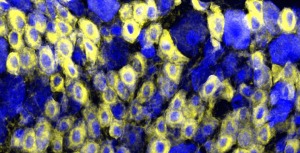
Prostatic acid phosphatase (PAP), seen here on the membranes of pain-sensing neurons (yellow), enduringly suppresses chronic pain. PAP could potentially provide long-lasting pain relief when administered before injury or inflammation.

“If you inject PAP before nerve injury or before causing inflammation, PAP has very long-lasting effect on the pain sensitization that follows,” said senior study author Zylka, assistant professor of cell and molecular physiology and a member of the UNC Neuroscience Center. “It has the potential to block or dramatically reduce pain, possibly in surgical settings.”

Zylka says PAP blocks pain in animal models by siphoning off a molecule called PIP2—a critical component of the chemical cascade behind chronic pain.

What’s more, PAP appears to keep on blocking pain symptoms long after it is injected.

Tens of millions of Americans suffer from chronic pain. This long-lasting pain is caused by a series of events along nerve cell membranes that make neurons hypersensitive. Injecting excess PAP into the system triggers a parallel series of reactions that makes it harder for this pain cascade to fire.

“Essentially PAP robs the cell of PIP2 so pro-pain pathways can’t signal as effectively.” explained Zylka. The team conducted their research using cell cultures and mice.

Using PAP to deplete PIP2 represents a promising new approach to treating chronic pain. “This is something people haven’t really focused on yet,” Zylka said. “We’re going right to the source of these pathways.”

In previous studies using mice, the team found that injecting PAP after an injury reduces sensitivity to both heat (like touching a hot burner) and mechanical sensitization (like the pain from brushing sunburned skin) for three days.

This time, the researchers took it a step further by injecting PAP before the injury. The effects lasted for the duration of the study—up to nine days.

Patients undergoing major surgery occasionally receive pain relievers through spinal injections just before the surgery begins. This study suggests that injecting PAP along with those other pain relievers might reduce patients’ need for analgesics like opiates in the days following surgery.  Future studies with patients will be needed to verify these possibilities.

“Ultimately, we’re very interested in other pain-related mechanisms that regulate PIP2 levels in cells. Any one of those mechanisms could be targeted for the treatment of chronic pain,” Zylka said.

Such research could provide new drugs for patients who already have chronic pain.

Les Lang and Anne Frances Johnson, science writer

Leave a comment

Filed under Uncategorized

Gut microbes, our extended family

Within the body of a healthy Homo sap adult, microbial cells are estimated to outnumber human cells by a factor of ten to one. The total number of genes associated with the human microbiome could exceed the total number of human genes by a factor of  100 to one.

So abundant among us  are these  fellow travelers that each of us should qualify for MFTN:  most- favored nation trading status. Or, at the very least, group health insurance.

These bacterial communities, complete with their own genomes, are still largely unstudied, leaving almost entirely unknown their influence upon human development, physiology, immunity, and nutrition.

In other words, it’s a good bet that we play host to a lot of good guys and bad guys.

Now a new UNC study  suggests that a shift in the balance between the “good” bacteria and the “bad” bacteria that populate our gut could be a harbinger of colon cancer.

The findings, which appear online in the May/June issue of the journal Gut Microbes, could lead to strategies to identify people who are at high risk as well as ways to manipulate the microbiota to prevent colon cancer.

Fluorescence in-situ hybridization (FISH) using bacterial 16S rRNA probes showing bacteria (in red)localized to the mucus layer directly on top of the crypts (yellow). Keku lab/UNC

“We think something happens to tip the balance away from the beneficial bacteria and in favor of microbes that make toxic metabolites and are detrimental to our health,” said senior study author Temitope Keku, Ph.D., research associate professor of medicine at UNC.

“By pinpointing these bacterial culprits, we can not only identify people at risk, but also suggest that they include the good bacteria in their diet,” added Keku. “And what a great way to address colon cancer – you could know your risk and lower it by eating your yogurt every day.”

Researchers have known for decades that the bacteria harbored in our bodies are not innocent bystanders but rather active participants in health and disease. Yet only recently have molecular methods evolved to the point that they can identify and characterize all of our microbial residents.

Keku and her colleagues used these methods to determine the different bacteria groups contained within biopsies from 45 patients undergoing colonoscopies. They uncovered a higher bacterial diversity and richness in individuals found to have adenomas than in those without these colorectal cancer precursors. In particular, a group called Proteobacteria was in higher abundance in cases than in controls, which was interesting considering that is the category where E. coli and some other common pathogens reside.

It’s still not clear whether alterations in bacterial composition cause adenomas, or if adenomas cause this altered balance.

In order to tell if it is the chicken or the egg, Keku plans to conduct more mechanistic studies, such as testing whether certain groups of bacteria promote cancer growth in animal models. She is also expanding the study to analyze samples from 600 patients using next-generation sequencing technology.

The ultimate goal may be to determine if the differences found in the mucosa lining the colon also exist in the fecal matter that passes through the colon. If so, it could mean less invasive screening for cancer and even more cancers being caught earlier, when survival rates are higher.

“We have come a long way from the time when we didn’t know our risk factors and how they impact our chances of getting colon cancer,” said Keku. “But now that we can look at bacteria and their role, it opens up a whole new world and gives us a better understanding of the entire gamut of factors involved in cancer – diet, environment, genes, and microbes.”

Les Lang and Marla Broadfoot

Leave a comment

Filed under Uncategorized

Schizophrenia brain signs in babies?

A long, long time ago,  in a galaxy just like ours….  

Let’s let’s try that again.

Some of you might recall a time when a popular idea regarding mental illness was that the fault, dear reader, was not in our genes  but in the way we communicated with each other.

In the  mid-1950s, a number of social scientists and psychotherapists developed proposals that attributed schizophrenia to the exposure to, and participation in, dysfunctional communication patterns in the family.

Some of the concepts flying around like drunken pelicans included  “double-bind communication,”  “pseudomutuality, and “pseudohostility.” (For a nifty overview, circa 1997, see this article.)

It was an intriguing idea that sort of jelled with a lot of us Homo saps who viewed mental illness as w-a-a-a-y more environmentally than genetically determined.  And even into the early 1960s, the majority of undergrad psych students would feel compelled to agree.

Today, in the nature versus nurture game, we  tend to be interactionists in our view of schizophrenia. We know it to be a debilitating mental disorder affecting one in 100 people worldwide, that most cases aren’t detected until a person starts experiencing symptoms like delusions and hallucinations as a teenager or adult. And by that time, the disease has often progressed so far that it can be difficult to treat. 

Infant’s brain image on left shows the larger lateral ventricles and a generally larger brain overall. Image provided by John Gilmore, MD.

 Now new research published online in the American Journal of Psychiatry offers the first evidence that early neonatal brain development may be abnormal in males at genetic risk fo schizophrenia.

 The scientists used ultrasound and MRI to examine brain development in 26 babies born to mothers with schizophrenia.  (Having a first-degree relative with the disease raises a person’s risk of schizophrenia to one in 10.)

 Among boys, the high-risk babies had larger brains and larger lateral ventricles—fluid-filled spaces in the brain—than babies of mothers with no psychiatric illness. The new findings were detectable in babies only a few weeks old.

“It allows us to start thinking about how we can identify kids at risk for schizophrenia very early and whether there things that we can do very early on to lessen the risk,” says lead study author John H. Gilmore, MD, professor of psychiatry and director of the  UNC Schizophrenia Research Center.

“Could it be that enlargement is an early marker of a brain that’s going to be different?” Gilmore speculates.

No difference was found in brain size among girls in the study. This fits the overall pattern of schizophrenia, which is more common, and often more severe, in males.

The findings, o course,  do not necessarily mean the boys with larger brains will develop schizophrenia. Relatives of people with schizophrenia sometimes have subtle brain abnormalities but exhibit few or no symptoms.

 “This is just the very beginning,” said Gilmore. “We’re following these children through childhood.”

 The team will continue to measure the children’s brains and will also track their language skills, motor skills and memory development. They will also continue to recruit women to the study to increase the sample size.

Les Lang

Leave a comment

Filed under Uncategorized

Autism genetics: an emerging consensus

A large international consortium of researchers, including scientists at the University of North Carolina at Chapel Hill, have announced new discoveries that could help clarify the genetics of autism.

Their findings published online June 9th in Nature,  support an emerging consensus among scientists that autism is caused by many “rare variants” or genetic changes found in less than one percent of the population.

While each of these variants may only account for a small fraction of autism cases, collectively they appear to account for a greater percentage of individuals within the autism community. They are also providing into possible mechanisms involved in the disease.

The Autism Genome Project (www.autismgenome.org) collected genotyping data from 1,000 individuals with autism spectrum disorder, or ASD, and 1,300 without ASD. They found that people with the disorder tend to carry submicroscopic insertions and deletions called copy number variants, or CNVs, in their genome. Some of these variants appear to be inherited, while others are considered new because they are found only in affected offspring and not their parents.

These CNV in individuals with ASD tend to disrupt genes previously reported to be associated with autism or intellectual disabilities.

The study also identified new genetic risk factors for autism (genes known scientifically as SHANK2, SYNGAP1, DLGAP2 and the X-linked DDX53–PTCHD1 locus.)

Some of these genes belong to nerve synapse-related pathways, while others are involved in cell proliferation, cellular movement, and intracellular signaling – functional targets that may lead to the development of new treatment approaches.

“These findings provide further evidence that autistic behavior is the result of many rare, small genetic changes,” said Joseph Piven, MD, study co-author and a lead AGP consortium investigator. Piven is also Sarah Graham Kenan Professor of Psychiatry at UNC and director of the Carolina Institute for Developmental Disabilities. 

“While genetic abnormality or relevant CNV identified appears to account for only a handful of affected individuals, taken together these various CNVs in different locations throughout the genome are beginning to account for a significant number of occurrences of autism in the population. Identifying the genes and biological pathways associated with these genes will eventually lead us to new treatments for autism based an understanding of the underlying biological causes.”

Geraldine Dawson, PhD, research professor of psychiatry at UNC, also coauthored the new study.

The AGP consists of 120 scientists from more than 60 institutions representing 11 countries who formed this first-of-its-kind autism genetics consortium.  Since 2002, group members have shared their samples, data, and expertise to facilitate the identification of autism susceptibility genes.

Les Lang

Leave a comment

Filed under Uncategorized

Good vibrations for male contraceptive

 Some might call it a kind of Holy Grail for reproductive science: the development of a long-term, inexpensive, completely reversible and nonhormonal male contraceptive, one that’s  suitable for use in developing to first world countries.

 And that is exactly the long-term goal that two UNC scientists have set out to accomplish. How? By using ultrasound from therapeutic instruments commonly found in sports medicine or physical therapy clinics. And their quest is well underway, with very promising results. 

James Tsuruta, PhD, assistant professor in the Laboratories for Reproductive Biology  in UNC’s Department of Pediatrics and Paul Dayton, PhD, associate professor and director of graduate studies in the Department of Biomedical Engineering successfully depleted testicular sperm in laboratory rats using therapeutic ultrasound. 

 And just this month, the pair received a $100,000 Grand Challenges Exploration grant from the Bill & Melinda Gates Foundation.  The project, aimed at further fine-tuning this technique for maximum effect and safety, could provide men with six months of reliable, low cost, non-hormonal contraception from a single round of treatment. 

Tom Hughes in our news office tells us that Tsuruta and Dayton’s project is one of 78 grants announced by the Gates Foundation in the fourth funding round of Grand Challenges Explorations, an initiative to help scientists around the world explore bold and largely unproven ways to improve health in developing countries.  The grants were provided to scientists in 18 countries on six continents.

The testis is composed of many tubes called “seminiferous tubules.”  In the accompanying image here, the seminiferous tubule on the left is from a testis that was not treated with ultrasound while the tubule on the right is from a testis that was treated with ultrasound. 

The tubule from the control testis has many darkly stained germ cell nuclei.  Most germ cell nuclei are round; the long, thin nuclei closest to the center of the tubule belong to germ cells called spermatids and they will soon be released as testicular sperm.  In contrast, the ultrasound-treated tubule is completely lacking testicular sperm and has lost almost all immature germ cells, decreasing its overall diameter while greatly increasing the amount of “empty” space in the center of the tubule.

“Once the testis has stopped producing sperm and all “sperm reserves” have been depleted, it is impossible to be fertile,” Tsuruta says. “Our Grand Challenges Exploration project will determine the appropriate ultrasound treatment to temporarily interrupt the supply of testicular sperm yet allow the testis to regenerate itself from the germ cells remaining after treatment.”

May the good vibes continue….

Les Lang

Leave a comment

Filed under Uncategorized

Palladin, the travels continue

Our second Hard Science blogpost “Have Palladin, Will Travel,” on January 19, 2009, introduced the work of UNC cell and molecular physiologist Carol Otey and the protein she discovered a decade or so ago that plays an important role in cell motility, adhesion and structure.

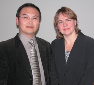
Study first author Silvia Goicoechea, PhD and, on the right, Carol Otey, PhD in the Otey Lab at UNC

Also mentioned was Otey’s most recent work in which  palladin was found mutated in an inherited form of pancreas cancer and was also produced in large amounts — “upregulated” — in a number of sporadic pancreas tumors.

Now it appears that the protein with the fancy name may prove to be a molecular marker of pancreatic cancer , one that might help spot the disease at its earliest stages, when it can be treated more successfully with surgery.

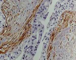
In a finding published April 26, 2010, in the online journal PLoS One, the researcher and her colleagues  showed that a specific form of a protein called palladin is produced in large amounts in the “tumor nest,” the cells that surround a pancreatic tumor. 

The blue cells in the middle, surrounding the “space” in the center of a pancreatic duct, are the tumor cells, and the brown cells around them are the tumor-associated fibroblasts (stained for palladin).

Measuring the levels of this form of palladin in patient samples could provide an improved way to screen for the deadly cancer, possibly catching it earlier than ever before, said senior study author Carol Otey, Ph.D., associate professor of cell and molecular physiology at UNC.

“The problem with pancreas cancer is it is almost never caught at an early stage,” said Otey. “By the time a person develops suspicious symptoms, the disease has typically progressed too far. But if you can diagnose it early, it can be treated very effectively with surgery.” 

Otey and her colleagues decided to see if the upregulation of palladin in the tumor nest could provide a useful diagnostic tool for identifying the disease at earlier stages. They knew that the single palladin gene message can actually be cut and pasted together in a manner that produces at least seven different palladin protein products. Turns out only a couple of these forms of palladin – called isoforms – appear in pancreatic tissue. 

The researchers found that the longer of these two isoforms was upregulated in the cells surrounding the tumor — called tumor-associated fibroblasts — when compared to normal pancreas. Their findings were consistent, regardless of whether they were looking in cultured cell lines, patient samples, or tumors from a mouse model. 

Otey thinks that the upregulation of this form of palladin in tumor-associated fibroblasts could help them become contractile and stiff — more like muscle than connective tissue — in order to generate channels through neighboring tissue so the cancer can metastasize and spread.  

“The interactions between these tumor-associated fibroblasts and tumor cells are really important and are probably what is causing pancreas cancer to be so deadly,invasive and resistant to current therapies,” said Otey.

And that is why raising public awareness and enhancing our abilities to diagnose the disease early is so critical, says Hong Jin Kim, associate professor of surgery at UNC, who along with Otey is senior author of the study.

“It appears that the upregulation of palladin in the tumor-associated fibroblasts is an early event in the neoplastic process,” said Kim. “We may be able to take advantage of these findings, since pathologic confirmation of pancreatic adenocarcinoma in the preoperative setting is often difficult, requiring an invasive procedure directed by endoscopic ultrasound.  If we can enhance the diagnostic efficiency of these studies by staining for palladin, it would be clinically helpful for interventional gastroenterologists and pathologists.”

For palladin, the travels continue….

Leave a comment

Filed under Uncategorized

Chemo drug awakens virus in cancer cells

Viruses and cancers interact in ways that were previously unknown  to science.  A new study led by UNC researchers shows that a common cancer drug can activate a viral infection that, paradoxically, can help anti-viral medications eradicate virus-associated cancer.  

The cooperative study, conducted by a team of UNC School of Medicine scientists and the UNC Project in Malawi, demonstrated for the first time in humans that a common drug used to treat Burkitt lymphoma can activate infection by the Epstein-Barr virus (EBV), a virus which typically lies latent inside the tumor cells of affected patients.

The finding paves the way for a future study using both a cancer drug and an antiviral agent to eradicate both the active virus infection and the tumor.  The study is reported in the April 1 issue of the journal Clinical Cancer Research.

Margaret L. Gulley, MD, professor of pathology and laboratory medicine, said,  “What we have learned from this work is a potential means of capitalizing on presence of viral genomes within tumor cells to alter those tumor cells in a way that makes them more susceptible to treatment. Our findings have implications for other EBV- related malignancies that, overall, are among the most common cancers worldwide.” Gulley is a member of UNC Lineberger Comprehensive Cancer Center. 

Weihua Tang, MD, PhD, the study's first author and research fellow with Margaret L. Gulley, MD

 EBV infects more than 90 percent of the world’s population and is associated with diseases ranging from infectious mononucleosis to lymphomas, gastric cancer and cancer of the nose and throat. 

Burkitt lymphoma, which is associated with EBV, is rare in most parts of the world, but is endemic in sub-Saharan Africa. Burkitt lymphoma is an aggressive, fast-growing type of non-Hodgkin lymphoma that often occurs in children. The disease may affect the jaw, bowel, lymph nodes, or other organs. 

The study demonstrated that initiating treatment with the anti-cancer drug  cyclophosphamide in children with Burkitt lymphoma simultaneously triggered an active EBV infection. The increased replication of EBV in cancer tissue makes these cells more susceptible to the antiviral drugs that kill cells containing replicating virus. Antiviral agents such as ganciclovir and valacyclovir are already in routine clinical use for treating active viral infections. 

Researchers enrolled 21 patients with a confirmed diagnosis of EBV-related Burkitt lymphoma. The patients ranged in age from 5-15 and were under treatment with cyclophosphamide for their cancer.  Through laboratory analysis of biopsy samples, researchers found that cyclophosphamide seems to induce the phase of viral infection most susceptible to antiviral therapy. 

Gulley points out that cyclophosphamide “is fairly efficacious, but it and even more expensive chemotherapeutic agents may be hard to come by in developing nations.”

Moreover, she says clinical trial data are needed to determine if chemotherapy and antiviral therapy synergize in managing Burkitt lymphoma.  

 “Our work provides scientific data to justify moving forward with a clinical trial.  Generic versions of several pertinent antiviral agents are available and are relatively inexpensive.”

Plans for such a trial are already underway under the leadership of Carol Shores, MD, PhD, associate professor of surgery in UNC’s Otolaryngology/Head and Neck Surgery Department and senior author of the study.  Shores says she is currently working to get approval for a Phase I/II clinical trial of the antiviral drug valacyclovir with cyclophosphamide in Malawi. “Valacyclovir recently went off patent, and a relatively generic form is available.  It is an oral drug approved in the US for children as young 3 years of age.

Leave a comment

Filed under Uncategorized